Saturday, September 30, 2023

Upper-Level Wenckebach

Here is another great ladder diagram from Chou's Electrocardiography in Clinical Practice (6 ed), Chapter 19: Atrioventricular Block; Concealed Conduction; Gap Phenomenon.


Incidentally, this hypothesis was constructed before the Kosowsky, et al, publication Multilevel Atrioventricular Block (Circulation, Vol 54, No 6, 1976).

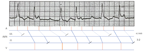
I wonder if we can further extrapolate the original diagram by subdividing the AV junction (or, AV node) into upper and lower level components. It may look something like this:


The basic idea is that there is Wenckebach conduction in the upper level (UL), and fixed ratio conduction in the lower level (LL). This can be tricky to appreciate, so the repeating pattern has been colorized for enhanced optics. 

The first cycle of the 4:3 Wenckebach (WB) is green, then repeats itself in yellow. I assume this pattern would continue to manifest had the telemetry been longer. 


The (assumed) LL fixed conduction ratio of 3:2 is demarcated in red and purple.


The opposite of this pattern (that is, fixed ratio in the UL and Wenckebach in the LL) is hypothesized here:

https://ecgladder.blogspot.com/2023/09/atrial-flutter-with-variable-block.html

Parasystolic Rhythm

This diagram is from Chou's Electrocardiography in Clinical Practice (6 ed), Chapter 19: Atrioventricular Block; Concealed Conduction; Gap Phenomenon.


(Please excuse the yellow highlighter.)

Notice how this masquerades as Mobitz II. The first clue to some other mechanism is the changing PR-interval related to QRS 2-4.


Peculiar P-QRS Interaction

 Another great case from Dave Richley, @DaveRichley


My hypothesis:


Orange arrows: Sinus P waves (mostly stable PP CL)

Solid black line: Fast Pathway (FP). Dashed black line: Slow Pathway (SP)


Fundamentally, dual AVN conduction. The basic, recurring them (demonstrated in QRS 2 and 3) Sinus conduction down FP. Then, preferential utilization of SP because FP is refractory to antegrade travels, however subsequently recovers enough to permit momentary retrograde conduction into the Atria (blue upright arrow). This would otherwise reset the PP CL, but the assumption here is that the SN is protected via entrance block. Thus, it fires, and the surrounding Atrial tissue is receptive to its impulse, however fails to effectively enter the AVN.


QRS 9 is ventricular extrasystole that invades the AVN and stifles what should have been SP conduction at this predictable moment in time. I initially considered the very next PR-interval (associated with QRS 10) to be the consequence of concealed conduction into FP. –And this remains a possibility. However, given that the PR-interval here shares similar distance to all previous SP conduction, I think it might be preferential use of SP because FP remains refractory from the extrastystolic penetration.


From Dave Richley:


In his words:


Beat 2: conducts down fast AVN p/way 


Beat 3: block in fast p/w. Conducts down slow p/w, then retrograde conduction up fast p/w & thru atria causing pseudo r’ and scooped ST seg. Retrograde concealed conduction from PVC blocks conduction in both p/ws.


AFib?

 ECG from Arron Pearce, @Arron_Pearce_


He asks:

Interesting rhythm case which I think is an atrial flutter with multilevel wenckebach block. I've never seen a baseline in lead II like it. I think it looks strange because of the way that the flutter waves are merging with the T waves Thoughts?


Dave Richley submitted this hypothesis:


Here's another depiction of multilevel AVN block:

https://ecgladder.blogspot.com/2023/09/atrial-flutter-with-variable-block.html


Complete Heart Block?

 This case is from the great ECG aficionado, Dave Richley. @DaveRichley


Regular QRS. Regular P waves. Are they dissociated?

His ladder diagram hypothesis:


In his words:

1. Atrial tachycardia (flat P waves I & V1; atrial rate 112/min). 

2. Wenckebach AV block and dual AVN pathways with conduction switching from fast to slow pathway explaining big jump in PR from beat 5 to 6 and beat 9 to 10.


Dual AVN Physiology

 Fantastic case from @AThomazAndrade


Here is Dr. Antonio's ladder diagram:


Read the full case report here:

https://jafib-ep.com/wp-content/uploads/2023/09/ECG-Challenge-Accelerated-Junctional-Rhythm-or.pdf


Ventricular Escape

 Twitter case from @ecgandrhythmRoe

 Seems to be some manner of escape rhythm, punctuated by Sinus capture.

 Hypothesis:

 The long horizontal lines demarcate zones of Atrium (A), AV node (AV), and Ventricles (V). 

The blue dots suggest a primary ventricular escape with successful retrograde conduction through the AV node, and into the Atrium. 

Suddenly, there is Sinus capture (green dot) with antegrade conduction into the ventricles. This Sinus impulse attempts to control the pace (orange dots), but is impeded because of restoration of the ventricular escape.


Atrial Flutter with Variable Block

 ECG posted to Twitter from @DanielSukmadja


Atrial Flutter with variable block. What sort of relationship is taking place between Atrium and Ventricles? My hypothesis is based on:

Multilevel Atrioventricular Block. Kosowsky, B. D., et al. Circulation, Vol 54, No 6, 1976.


The AV node (AVN) is physiologically divided into upper-level (UL) and lower-level (LL) components, each of which can influence antegrade conduction from Atrium to Ventricles.

I conceptualize it like this: 


Each colorized arrow is an antegrade impulse. The green impulse is blocked at the UL. The orange arrow is blocked at the LL. The black arrow successfully reaches the ventricles. Sometimes this behavior, collectively, manifests in a predictable pattern.


The alternating green and blue is to suggest that the UL has a 2:1 block. So, for every 2 impulses that enter the UL, 1 is blocked and does not proceed.


The alternating yellow and orange is to suggest that the LL has a 3:2 block. For every 3 impulses that enter the LL, only 2 proceed into the ventricles. Moreover, the shaded secondary impulse is to suggest that, at this level, the block is particularly Wenckebach.


Wide and Narrow QRS

This ladder diagram is from the following publication Atrioventricular Block with Narrow and Wide QRS: The Pause That Refreshes . The full a...